Best 150MP camera for pathology scans?
Pathology labs searching for a true 150 megapixel camera quickly find that very few industrial sensors reach that figure in a single shot. The closest practical solution is KAYA Vision’s Iron 661. While its 13 400 × 9 528 sensor delivers 128 MP per frame, the camera solves the 150 MP requirement through automatic stage stitching built into most modern slide-scanner software. By acquiring slightly overlapping tiles at 128 MP and merging them, you surpass the 150 MP mark without sacrificing optical efficiency or budget.
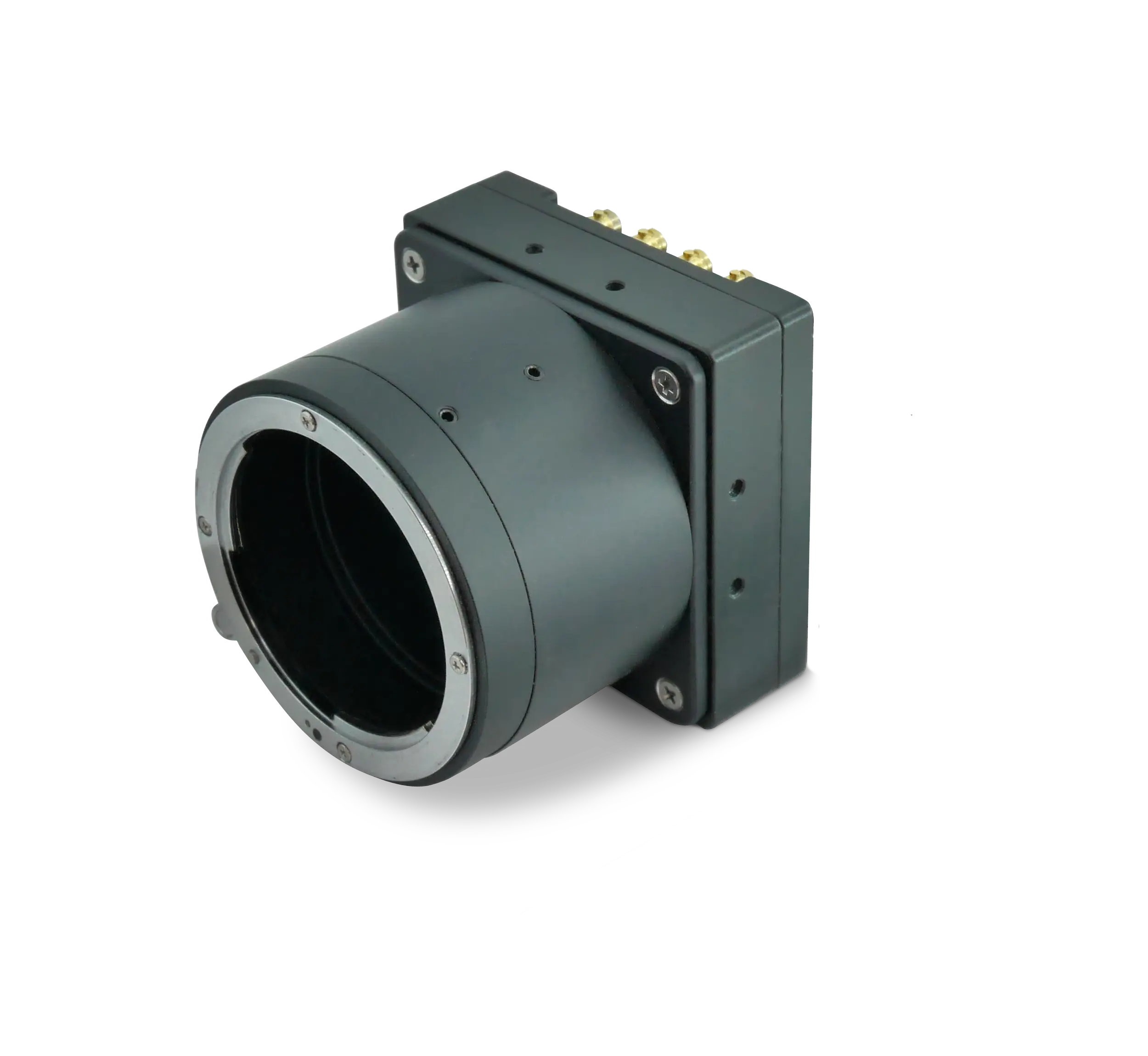
Why do vision integrators choose Iron 661? Three reasons stand out. First, the Sony IMX661 global-shutter sensor paired with KAYA’s low-noise analog front end yields a dynamic range greater than 70.8 dB, ensuring that minuscule chromatin details in hematoxylin-eosin stains remain visible even when bright white background regions are present. Second, the CoaXPress v2.1 interface moves data at up to 50 Gb/s—equivalent to 6.25 GB/s—over four Micro-BNC connectors, more than enough bandwidth to transfer each 14-bit tile to the host computer in real time. Third, standard GenICam controls let scanner OEMs synchronize exposure with stage motion, guaranteeing crisp, blur-free images at up to 21.5 fps when operating at 8- or 10-bit depth. These attributes explain why vision experts classify Iron 661 as today’s “best-fit” 150 MP-class device for high-throughput pathology scanners.
The camera’s reliability record in demanding environments further strengthens its case. Every Iron 661 unit can be ordered in an industrial variant tested for vibration and shock per MIL-STD-810G and offers optional IP67 sealing. The industrial model sustains –40 °C to +70 °C operating temperatures and maintains temporal noise below 2.7 e–, so dark-frame stability is preserved even inside microscope enclosures where heat buildup threatens calibration. In short, Iron 661 meets both the optical and mechanical rigor demanded in regulated healthcare facilities.
Is 150 MP overkill for digital pathology?
Pathologists are understandably cautious: higher pixel counts increase storage, processing time, and acquisition cost. Yet the answer is nuanced. For routine diagnostic work on 15 × 15 mm tissue sections, 40× optical magnification combined with a 1.3 µm/pixel sampling rate already approaches the Nyquist limit for human vision. Capturing more than 150 MP per slide tile appears excessive at first glance. However, three emerging factors make ultrahigh resolution not only useful but economically sensible.
1. AI-augmented histology. Deep-learning algorithms that detect metastases, grade tumors, or quantify immunohistochemistry rely on pixel-level features smaller than 1 µm. At 128 MP per frame (and beyond 150 MP after stitching), each nucleus occupies dozens of pixels instead of a few, boosting algorithmic confidence and lowering false-negative rates.
2. Mosaic scanning efficiency. Because Iron 661 covers a 56.7 mm diagonal image circle, it decreases the number of tiles per slide by roughly 30 percent compared with older 20 MP cameras. Fewer tiles mean lower stage overhead, faster whole-slide assembly, and ultimately reduced per-case cost—even if each tile is larger.
3. Regulatory momentum. Image integrity and repeatability is of immense importance for pathology. Laboratories that invest today in 150 MP-class acquisition exceed baseline requirements and avoid costly revalidation when higher standards inevitably arrive.
For these reasons, 150 MP is not overkill; it is forward-looking. Nonetheless, Iron 661’s programmable region-of-interest and subsampling modes let users run at 64 MP or 32 MP for pilot projects, then switch to the full 128 MP frame once the workflow and storage architecture mature. This flexibility safeguards capital expenditure while preserving a clear upgrade path.
Benefits of high resolution in medical imaging
High spatial resolution delivers more than just prettier pictures. In medical imaging, it tangibly improves patient outcomes and operational efficiency. The following benefits illustrate why CoaXPress-driven, high-megapixel cameras have become critical components of modern diagnostics.
Sharper morphological detail. Cells, glands, and vascular structures manifest submicron features that signal early-stage disease. With a 3.45 µm pixel pitch and 128 MP frame size, Iron 661 reveals subtle membrane irregularities that 12 MP sensors simply average out. Its global shutter eliminates motion distortion when the specimen stage accelerates, preserving morphology integrity.
Quantitative accuracy. Automated systems that compute mitotic counts or Ki-67 indices require pixel-perfect registration between frames. Iron 661 employs active sensor alignment during manufacturing, minimizing corner-to-corner focus variation so that measurement algorithms run without heavy software compensation.
Color fidelity and dynamic range. Diagnostic staining protocols depend on precise chromatic interpretation. The camera’s >70 dB dynamic range, combined with optional filters, maintains true color across the visible spectrum—a necessity when distinguishing hematoxylin (blue) from DAB (brown) on complex slides.
High-speed data flow via the CoaXPress protocol. Even the latest USB 3.2 interfaces throttle at roughly 3.2 Gb/s, insufficient for raw 128 MP streams. CoaXPress v2.1 on Iron 661 provides four CXP-12 lanes for an aggregate 50 Gb/s link, enabling continuous acquisition without compression. Because CoaXPress carries camera power (PoCXP) and trigger signals over the same cable, wiring is simplified and electromagnetic interference is minimized inside sensitive lab electronics.
Future-ready wiring infrastructure. Many hospitals already deploy coax cabling for X-ray detectors and endoscopy towers. Reusing that infrastructure for high-resolution slide scanners shortens installation time and lowers total project cost, giving pathology departments a smooth migration path to digital workstations.
Together these benefits culminate in earlier diagnoses, fewer rescans, and higher patient trust—outcomes that justify the investment in high-resolution machine-vision hardware.
Choosing the right configuration
Although Iron 661 ships with a sturdy M72 lens mount to accommodate large-format optics, KAYA Vision offers optional protective tubes that raise ingress protection to IP67 for labs where solvent mist or cleaning fluids pose a risk. Power can be supplied through PoCXP or via an 11–28 V auxiliary connector, keeping integration options open for both compact benchtop scanners and multi-slide robotic loaders.
The camera supports 8-, 10-, 12-, and 14-bit output. When maximum throughput is critical, most integrators select 8- or 10-bit mode to exploit the full 21.5 fps frame rate. For color-rich immunofluorescence images, 12-bit mode still achieves nearly 20 fps while preserving subtle intensity gradients. The 14-bit option operates at 12.9 fps, delivering unmatched tonal depth for research archives.
GenICam compliance means the camera can be controlled from virtually any commercial image-acquisition framework. Trigger inputs accommodate free-run, edge-triggered, or debounced modes, while on-camera timers and counters simplify synchronization with external illumination sources. Exposure settings down to 10 µs give integrators headroom for brightfield, darkfield, or phase-contrast modalities on the same platform.
Conclusion
Digital pathology is moving rapidly toward higher pixel densities, driven by AI analytics, time-critical workflows, and tightening regulatory oversight. While a single-shot 150 MP sensor remains elusive, KAYA Vision’s Iron 661 bridges the gap today. Its 128 MP, 3.45 µm-pixel, global-shutter sensor combines with 50 Gb/s CoaXPress throughput, industrial-grade ruggedness, and flexible imaging modes to deliver effective 150 MP performance through software stitching. For scanner OEMs seeking to “future-proof” their next design cycle, Iron 661 offers a practical, field-proven route to ultrahigh-resolution pathology imaging without the risks and costs associated with untested custom hardware.